- By JRSH Admin
- In Health and Tips,
- Posted August 26, 2021
Spina Bifida: Causes, Symptoms, Types, and Treatment
What is spina bifida?
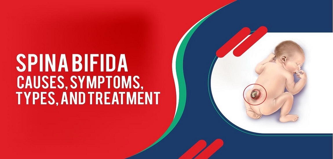
Spina bifida, a spinal condition that affects newborns at birth, is generally detectable. It is a common type of neural tube defect (NTD). The spinal cord and spine do not develop properly in babies with spina bifida, which causes birth defects. In an embryo, the neural tube develops into the brain, the spinal cord, and the tissues that surround them.
An infant with the condition will not form a fully developed spine if their neural tube (a group of cells that forms the brain and spinal cord) doesn't completely close. These conditions can lead to physical and mental health problems.
A cyst can be found anywhere along the spine and is usually visible in an opening in the back of the baby at birth. It can also appear as a sack of fluid growing on the spine outside the body.
What is the prevalence of spina bifida?
Spina bifida is only detected in 1-2 out of every 1,000 live births globally. In India, this number ranges from 4 to 8 out of every 1,000 live births, depending on the state.
It is believed that this is due to a lack of awareness regarding the need for folic acid during the first trimester of pregnancy.
With vitamin and folic acid, approximately 72% of spina bifida and neural tube defects (NTDs) can be prevented.
Types of Spina bifida
There are three types of spina bifida i.e. Spina bifida occulta, Meningocele, and Myelomeningocele.
1. Spina bifida occulta:
Among spina bifida types, occulta is the mildest. This condition is sometimes called “hidden” spina bifida. As a result, you have a small opening in the spine, but no sac whatsoever on your back. Nerves and the spinal cord are usually normal. In most cases, spina bifida occulta does not manifest itself until late childhood or adulthood. It typically occurs at the levels of the 5th lumbar and 1st sacral vertebrae. Unlike some other types of spina bifida, this one usually does not cause any disabilities.
2. Meningocele:
Associated with this type of spina bifida is a sack of fluid on the back of the baby. Fortunately, the sack does not have any spinal cord in it. Meningocele causes only minor disabilities as there is not much nerve damage. Most people do not have symptoms, but some have problems with their bladder or bowels.
3. Myelomeningocele:
One of the most severe forms is myelomeningocele, also known as open spina bifida. There are several vertebrae in the lower back or middle back that are open to the spinal canal. When the membranes and nervous system emerge through this opening at birth, a sac is formed on the baby's back, usually revealing tissues and nerves. Spina bifida of this type causes moderate to severe disabilities. A baby may suffer life-threatening infections as well as paralysis and bladder and bowel problems due to this.
Causes of spina bifida
Genetics and environment may both contribute to the development of spina bifida. A child affected by the condition, or a parent who has the condition, will have a 4% likelihood of having another child affected. Other risk factors include chronic seizures, folate deficiency, obesity, and poorly controlled diabetes. A higher risk exists for people who are white or Hispanic. Generally, girls are more likely than boys to suffer from spina bifida.
- Folate deficiency. Vitamin B-9, folate, plays a significant role in the development of a baby's health. In supplements and fortified foods, folic acid is found in a synthetic form. Spina bifida and other defects of the neural tube are linked to folate deficiency. Due to its water-soluble nature, folic acid does not stay in the body for long, so it has to be taken every day for it to be effective on neural defects. Researchers have found that by taking a multivitamin containing folic acid, women of childbearing age would have a 70% lower risk of neural tube defects.
- Family history of neural tube defects. The chance of a second child having the same defect is slightly higher for couples who have had one child with the defect. If both of your previous children had the condition, your risk is increased. In addition, women born with neural tube defects are more likely to give birth to children with spina bifida. Despite this, children with spina bifida are generally born to parents with no previous family history of the disease.
- Some medications. During pregnancy, anti-seizure medications, including valproic acid (Depakene), can cause neural tube defects. The body may have trouble using folate and folic acid because they interfere with the process.
- Diabetes. The risk of having spina bifida is higher in women with diabetes who have poorly controlled blood sugar.
- Obesity. A woman who is obese pre-pregnancy is more likely to give birth to a child with neural tube defects, such as spina bifida.
- Alcohol. Misuse of alcohol can lead to macrocytosis, which destroys folate. For bone marrow to rejuvenate and recover from macrocytosis, it needs months after the consumption of alcohol is discontinued.
- Increased body temperature. According to certain studies, excessive body temperature (hyperthermia) in early pregnancy may increase the risk of spina bifida. The risk of spina bifida may be slightly increased when your core body temperature is elevated due to fever or the use of a sauna or hot tub.
Symptoms of spina bifida
Symptoms of spina bifida occulta include:
- There is a space between vertebrae
- The exterior does not reveal any openings
- External fluid-filled sacks are not present
- Several birthmarks or dimples on the back
- A small cluster or group of hair on the back
- Fat deposit on the back
Symptoms of meningocele spina bifida include:
- An opening can be seen in the back
- At birth, a sack is visible
- Via the vertebral opening, the membranes push into the sack
- It is normal for the spinal cord to develop
Symptoms of myelomeningocele spina bifida include:
- Some vertebrae have an open spinal canal, usually in the middle or lower back
- An exposed sack or sack covered with skin that contains the spinal cord and membranes
- Muscle weakness or paralysis in the legs
- seizures
- deformed feet
- hips that are not even
- scoliosis (curved spine)
- issues with the bowel and bladder
Diagnosis of Spina Bifida
Your pregnancy may be screened for spina bifida and other birth defects during prenatal testing. Testing isn't perfect. Spina bifida may not be present in all babies of mothers who have positive blood tests. There remains a small possibility of spina bifida even if the test results are negative. Prenatal testing can have risks, so speak with your doctor about how you might cope with the results.
Blood tests:
Maternal blood tests can detect spina bifida, but ultrasound is normally used to diagnose it.
- Maternal serum alpha-fetoprotein (MSAFP) test. MSAFP involves taking a sample of the mother's blood and testing it for alpha-fetoprotein (AFP) - a protein produced by the baby. There is a possibility that some AFP will cross the placenta and enter the mother's bloodstream. AFP levels that are abnormally high indicate a neural tube defect such as spina bifida, though high AFP levels are not always associated with this condition.
- Test to confirm high AFP levels. Your doctor may order a follow-up blood test for confirmation if your AFP levels change due to other factors, such as an incorrect fetal age estimate or multiple births. A further evaluation, including an ultrasound exam, will be required if the results are still high.
- Other blood tests. The MSAFP test may be performed with two or three other blood tests. Together with the MSAFP test, these tests examine other abnormalities, such as trisomy 21 (Down syndrome), rather than neural tube defects.
Ultrasound:
Spina bifida can be detected by fetal ultrasound before birth with the greatest degree of accuracy. It is possible to perform an ultrasound in the first trimester (11 to 14 weeks) and the second trimester (18 to 22 weeks). Second-trimester ultrasound scans can identify spina bifida accurately. A congenital anomaly like spina bifida must be ruled out during this examination.
Spina bifida can also be detected by advanced ultrasounds, such as a wide spine or certain features of your baby's brain indicative of spina bifida. Ultrasound can also be used to assess severity when performed by an expert.
Amniocentesis:
Your doctor may request an amniocentesis if the prenatal ultrasound confirms the diagnosis of spina bifida. As part of amniocentesis, the doctor removes a sample of amniotic fluid from the baby's surrounding sac using a needle.
While spina bifida is not often associated with genetic diseases, this examination may be necessary to exclude them.
Diagnosis after the Baby Is Born:
In some cases, spina bifida cannot be detected until after the baby is born. It is not uncommon for a baby's back to have a hairy patch or a dimple that is visible shortly after birth. Imaging tests, such as a CT scan, MRI, or X-ray, can help doctors see the spine and the bones inside the baby's back with greater clarity.
When spina bifida is not diagnosed until after birth due to prenatal care not being received by the mother or incomplete ultrasound images showing the affected part of the spine, it is often too late for diagnosis.
Treatment of Spina bifida
Fetal Surgery:
In a randomized study (MOMS Trial), repair of the defect during pregnancy was compared to repair after birth, and the results were encouraging but mixed. There was a significant difference between patients who underwent fetal repair and those who did not (44 vs. 84%) and a difference in walking ability (44 vs. 24%). In addition, the fetal surgery group experienced a greater number of complications for both mother and child. According to the American College of Obstetricians and Gynecologists (ACOG), fetal surgery is only considered at centers with a team that has experience with fetal surgery owing to the higher risk of complications and the lack of long-term follow-up.
Infant Surgery( surgery after birth):
To prevent infection and further injury to the spinal cord, a baby born with spina bifida must have the exposed spinal cord repaired. They then close the muscles and skin after placing the neuronal tissues back in the spinal canal. In cases where there is a large opening that is hard to close, a plastic surgeon may get involved. The procedure is normally performed a few hours after birth as a medical emergency. Typically, the surgery is performed within 48 hours after the baby's birth.
Approximately 80-90% of spina bifida children develop hydrocephalus. Hydrocephalus is characterized by excessive cerebrospinal fluid (CSF) accumulating in the brain ventricles (fluid-filled cavities), which can result in increased head pressure. To control the build-up of spinal fluid, most of these children will require a ventricular shunt. Shunts are usually left in place for the duration of the individual's life, but they need to be replaced several times.
Prevention from Spina Bifida
- Pre-conception folic acid intake of 400 micrograms (mcg) a day can reduce the risk of spina bifida in children. Fruits, whole grains, fortified cereals, dried beans, leafy vegetables, and beans are all good sources of folic acid. Folic acid must be taken by all women, whether or not they intend to become pregnant since half of all pregnancies are unplanned. Researchers have found that by taking a multivitamin containing folic acid, women of childbearing age would have a 70% lower risk of neural tube defects.
- Discuss any medications, vitamins, dietary supplements, and over-the-counter drugs you are taking with your doctor or pharmacist.
- Ensure that your medical conditions are under control before you decide to get pregnant, such as diabetes and obesity.
- Make sure your body is not overheated, as would be the case if you used a hot tub or a sauna.
Tags
Blog Search
Latest Posts
-
Arthroscopy Surgery: Procedure, Benefits, and Recovery
July 04, 2025 -
What Are the Symptoms, Causes and Treatments of High Cortisol Levels?
June 16, 2025 -
7-Day Diet Plan For Diabetic Patients
May 16, 2025 -
What Is the Normal Sugar Level After Meals & How to Lower It Quickly with Food?
May 09, 2025 -
Understanding Nursing care plan for Asthma ( NCP for Asthma )
April 22, 2025